Histology: is the study of tissues
Tissue: is a group of similar cells combined into a specialized manner and preform a particular function. A tissue contains intercellular substances between cells, and contains tissue fluid surrounding the cell.
- Tissues vary greatly in appearance and structure
- Examples of tissues are: hard bone, sturdy/elastic skin, lining of respiratory tract, salivatory glands, blood, and soft muscle.
Components of Tissue
1. Cells: Differ in size, shape and structure. Ex: red blood cells (have no nucleus), white blood cells (one or several nuclei), muscle cells (contract), fat (have a displaced nucleus)
2. Intercelluar Substance: in between cells in all tissue of the body. It is a by-product of the cell. There are two main forms 1) Fibrous or thread, 2) Amorphous (without form/unicellular)
3. Tissue Fluid: part of the blood plasma which can diffuse thought the capillary wall (nutrients and wastes). Different tissue have different amount of tissue fluid. Ex: blood has a large tissue fluid, while skin has a small tissue fluid.
Human tissues are classified into four primary groups.
1. Epithelial tissue: is skin, it occurs as covering or lining membranes. All epithelial tissue rests on connective tissue, but are not alike in shape and arrangement. The major groups of epithelial tissue are surface cells of covering and lining membrane, and glandular tissue.
Shape :
- Squamous = flat
- Cubodial = cube
- Columnar = column
- Transitional = changes shape
- Simple: a single layer
- Stratified: several layers
- Pseudstratisfied: appears to be several layers but its actually only one.
2. Connective tissue differs greatly in form and function, and all lining of the epithelial and glandular tissue resides on the connective tissue. There are six types of connective tissue: fibours, loose alveolar, aclipose, hemopoetic, cartilage and bone.
- Specialized connective tissue are fat displaced nucleus', hemopoetic=blood, cartilage, and bone.
3. Nerve tissue: have cells that make up the nervous system are neurons. The central nervous system include the brain and spinal cord (no connective tissue)
4. Muscle tissues: there are three kinds of muscle tissue
- Smooth muscle: non striated, walls of blood vessels (involuntary control)
- Skeletal muscle: striated, very strong (voluntary control)
- Cardiac muscle: appear striated or striped when magnified (involuntary control)
When we are just forming in the womb, we are just two tubes. Larger tube is the outer body wall, the smaller inner tube is the digestive tract. In between the tubes are our internal organs. At either end of the tube is the mouth, and the other your butt.

This is basically what we all look like at 3 weeks old.
There are primary embryonic layers that our mouth, bone, tongue, lips, muscles etc, come from. All skin, tooth enamel, nose, and nervous system come from the ectoderm. The bones, and muscles come from the mesoderm, and the stomach, intestines and lungs come from the endoderm.
The Development of the Face
- Primitive mouth (stomodeum, which looks like the baby's forehead in the picture) is the clefting or invagination of the ectoderm and will eventually become the oral and nasal cavities
- Buccopharyngeal membrane (below the primitive mouth, looks like the back of the mouth) is where the endoderm and the ectoderm contact. It will eventually form the the palate and tonsil area. It will create an opening to the mouth; the membrane ruptures at week four with stomodeum to create upper end of the future gut.
- Rathkes Pouch (behind the buccopharyngeal membrane) is an extension of the buccophrygeal membrane before rupture. It meets downwards from the brain. It becomes the future pituitary gland which controls growth and development.
- Brachial Arch 1 ( literally looks like a little chin, 5 in total) is located below the stomodeum, and begin developing in the fourth week. It develops the oral and nasal cavity.
- Frontal Process is above the stomodeum, and will be the future forehead, nasal septum, and front part of the roof of your mouth.
Note: Most of the face is all developed from the branchial arch except for the tongue. The oral and nasal cavity are formed from both the frontal process and the first branchial arch.
Development of the Nasal Cavity
- It develops in the fourth week
- Invagination of the frontal process forming nasal pits
- these divide into: median nasal process and the left and right lateral nasal process
Median Nasal Process: grows down into the globular process which creates the tip of your nose and septum (for median nasal process), and your pituitary gland and premaxilla (upper mouth) for the globular process.
Lateral Nasal Process (left and right): grows the left and right side of the nose
Development of the Cheeks and Lips
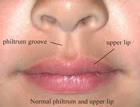
Maxillary Process (right and left) comes from branchial arch 1, creating the left and right cheek and left and right lip. It will grow into the mid line of the face and fuse with the globular process creating the philtrum and form the upper lip (sixth week)
Development of the Palate
Palatine Process (left and right): comes from the left and right maxillary process forming the right and left palate
Development of the Lower Part of the Mouth
Mandibular Process comes from the branchial arch 1 to form the mandible (lower jaw including teeth), parts of the front part of the tongue and the lower part of the face.
The rest of the tongue comes from branchial arch II forming the base of the tongue (down the throat)
The clinical significance of knowing how exactly all these process comes together and how they form is to understand incorrect growth/fusion of the processes. Some examples of incorrect growth and fusion of the processes is a cleft lip, cleft palate or an epithelial rest.
Cleft lips are a failure of the first or both maxillary processes with the globular process.
Cleft palate is formed by the failure of the fusion of anterior palate and a failure of the right and left lateral palatine processes to fuse with the premaxillary area. It can occur with or without a cleft lip
- Mildest form: the uvula is split in half
- Moderate form: soft palate and some of hard palate is cut in half
- Sever form: lack of fusion with the bone that supports the teeth (alveolar bone)


Treatment
- Cleft lip is easier to repair, as early as 12 weeks old.
- Cleft palate usually 18 months depending on the extend of bone loss.
- an applance may be required to remove the gap from mouth to nose
- speech therapy is usually required
Cleft lip is more common in boys
5500 case per year
Cleft palate is more common in girls
Epithieal Rest
Is a misplace clumps/groups of epithelial cells. They are loged between embryonic processes that are coming together. These cells come from the ectoderm, and they cause a formation of a cyst
- Odontoma: where something goes wrong in the tooth cells
- Branchial Cleft Cyst: swelling in the neck.


No comments:
Post a Comment